Introduction
(Pneumothorax)
neumothorax refers to the presence of air or gas in the pleural cavity between the visceral and parietal pleura, which results in violation of the pleural space. It is uncommon during childhood, but can be life threatening. Primary spontaneous pneumothorax occurs in children without known lung disease, whereas secondary spontaneous pneumothorax occurs as a complication of chronic or acute lung disease. Traumatic pneumothorax is caused by blunt or penetrating trauma to the chest. Iatrogenic pneumothorax is a complication of certain diagnostic or therapeutic procedures such as central line placement or as a consequence of mechanical ventilation.
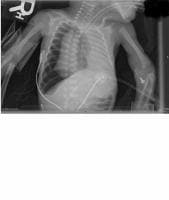
The image below depicts a radiograph of a neonate with pneumothorax.
Neonate with a right tension pneumothorax. Note the tracheal deviation to the left.
Pathophysiology
Pneumothorax can be classified as either simple or complicated. Spontaneous pneumothorax occurs via rupture of the visceral pleura, whereas traumatic pneumothorax may occur following injury to either pleural layer. In both types, a loss of intrapleural negative pressure causes lung collapse.
The main physiologic consequences of a pneumothorax are a decrease in vital capacity and a decrease in PaO2. Most patients with a pneumothorax have a reduced PaO2 and an increased alveolar-arterial oxygen tension difference. The reduction in PaO2 appears to be caused by areas with low ventilation-perfusion ratios, anatomic shunts, and alveolar hypoventilation.
In a simple pneumothorax, air in the pleural space does not build up significant pressure but allows the lung to collapse 10-30% without further expansion of the pneumothorax. A small pneumothorax may be asymptomatic and well tolerated.
A complicated pneumothorax is progressive and consists of continued air leakage into the pleural space that can not exit during exhalation. This results in progressive lung collapse. This continued air leak results in positive pressure within the hemithorax and displacement of the mediastinum (ie, tension pneumothorax).
Tension pneumothorax is a life-threatening emergency. The positive pressure results in collapse of the involved lung and a shift of the mediastinal structures to the contralateral side. This causes a decrease in cardiac output as a consequence of decreased venous return and leads to rapidly progressive shock and death if not treated.
Frequency
United States
The annual incidence of primary spontaneous pneumothorax in the general population is estimated to be 5-10 per 100,000 population. The peak incidence occurs in persons aged 16-24 years. The disorder is less common in children than in adults.1 The rate of pneumothorax is relatively higher in the newborn period, even in full-term newborns, but declines during infancy.2
Sex
Limited data in young children suggest a strong male predominance of primary spontaneous pneumothorax.
Age
All age groups are affected. Premature neonates on mechanical ventilation are at high risk.
Clinical
History
Spontaneous pneumothorax often occurs when a patient is at rest or with minimal exertion. Patients who are symptomatic may complain of the sudden onset of pleuritic chest pain that is sharp or stabbing and may be preceded by a popping sensation. Guidelines for the differential diagnosis of chest pain are available.3 These patients may also have sudden onset of dyspnea. Patients with small pneumothoraces may occasionally have a dry or nonproductive cough.
The evaluation of patients presenting with a spontaneous pneumothorax should always include the investigation of potential causes such as use of inhaled drugs, asthma, foreign body aspiration, infections, and connective tissue diseases.
Physical
The severity of the symptoms depends on the extent of the lung collapse, rate of development, and underlying clinical status of the patient.
A patient with a simple pneumothorax may either be asymptomatic or present with symptoms such as chest pain and dyspnea. Patients with a spontaneous pneumothorax secondary to underlying lung disease may have a more dramatic presentation.
A more extensive pneumothorax often produces pleuritic chest pain, dyspnea, tachypnea, cyanosis, and decreased breath sounds on the involved side. Acute pleuritic chest pain on the affected side occurs 95% of the time.
Hyperresonance to percussion may be noted on the affected side. Subcutaneous emphysema with crepitance is occasionally present.
Patients with a tension pneumothorax may present in shock and have trachea displacement to the unaffected side.
If the pneumothorax is due to trauma, look for contusions or abrasions on the chest wall or a small puncture wound that does not allow free movement of air between the outside and the pleural cavity.
Causes
Simple or complicated pneumothorax is very common in both blunt (38%) and penetrating (64%) pediatric chest injuries.
Cases not associated with trauma are generally due to a pulmonary bleb rupture with subsequent air leakage into the pleural space.
Inhalation of some toxic substances, most notably crack cocaine, can also lead to this condition.
Spontaneous secondary pneumothoraces may occur in patients with underlying lung diseases such as asthma, cystic fibrosis,4 or pneumonia.
When trauma results in pneumothorax, it may be secondary to blunt trauma or penetrating trauma. Penetrating trauma results in open or communicating pneumothorax.
Workup
Laboratory Studies
Patients who present with respiratory distress should have an ABG assessment.
Hypoxemia occurs because of significant ventilation perfusion mismatch.
Hypercapnia is unusual in patients without underlying lung disease.
Imaging Studies
Pneumothorax is generally a clinical diagnosis that is confirmed with upright chest radiography. Anteroposterior and lateral views can reveal the presence of even small amounts of intrapleural air. Air in the pleural space that outlines the visceral pleura is a characteristic finding. Hyperlucency of vascular and lung markings on the affected side can be seen because of this air. Atelectasis may also be seen on the affected side, and the mediastinum and trachea may shift away from the pneumothorax.
A small pneumothorax in a supine patient can be more easily detected in the lateral decubitus view. A noncontrast chest CT scan may be helpful to look for preexisting pulmonary pathologies such as blebs or bullae. Ultrasonography has also shown to be useful in detecting pneumothorax.5
A tension pneumothorax should always be a clinical diagnosis because death can occur before the radiograph is obtained or developed.
When an infant is suspected of having a pneumothorax, anterior-posterior radiographs are obtained in the supine position. Small pneumothoraces can be better visualized with lateral decubitus film with the affected side up.
Other Tests
Transillumination of the chest may help to establish the diagnosis in the newborn infant.
Treatment
Medical Care
In general, treat a small (<25%),>
A small, simple pneumothorax in a patient who experienced trauma is best treated with a chest tube because it may rapidly convert into a tension pneumothorax, especially if positive pressure ventilation is applied. Large or significantly symptomatic pneumothoraces require chest tube placement and surgical intervention. A tension pneumothorax requires immediate decompression with needle thoracostomy.
Patients with cystic fibrosis who sustain recurrent pneumothoraces may benefit from sclerotherapy, although this may not prevent all future recurrences.
Surgical Care
Percutaneous aspiration or tube thoracostomy (chest tube) placement is typically required for large or symptomatic pneumothoraces. Tension pneumothoraces need immediate decompression with needle thoracostomy, followed by tube thoracostomy.
Only pleurodesis (intrapleural instillation of a sclerosing agents such as talc, fibrin glue, and antibiotics), video-assisted thoracoscopy (VATS), and thoracotomy reduce the risk of future recurrence. Pleurodesis may be indicated for the first episode of a secondary pneumothorax due to the high incidence of recurrence or after a recurrence of a primary pneumothorax.6
Consultations
Unless the physician is skilled at placing a chest tube and handling potential complications, an emergent emergency medicine, pulmonary, critical care, or surgical consultation is warranted.
Diet
For patients with small pneumothoraces that are being watched, restriction to nothing by mouth (NPO) or clear liquids is appropriate. If chest tube placement is imminent, the patient should be NPO. After chest tube placement and recovery from any sedation, a regular diet may be started unless contraindicated based on other injuries or planned surgery.
Activity
A patient with a chest tube in place may only be able to tolerate restricted movement, such as from a bed to a chair.
Medication
The only drug therapy that is currently a component of the standard of care for pneumothorax is administration of 100% oxygen. Patients with a thoracostomy tube in place should receive appropriate pain management. Patients in severe pain should receive morphine sulfate intravenously or a patient-controlled analgesia pump when appropriate. Some patients' pain can be controlled well with oral medications such as acetaminophen.
Further Inpatient Care
After a small pneumothorax has resorbed completely, the patient requires no further inpatient care.
If a chest tube was placed, daily radiographs are sometimes required. If reexpansion occurred on initial placement with no subsequent air leaks, the tube can be removed in as little as 2-3 days. The decision to remove a chest tube is generally made by the surgeon, not the pediatrician.
Further Outpatient Care
Instruct parents to return if the patient has chest pain or shortness of breath.
The chest tube wound site should be monitored for infection and to ensure proper healing.
Inpatient & Outpatient Medications
Pain medication should be given on discharge.
Transfer
If the patient is in a clinic or office setting, order an immediate transfer to an emergency department by ambulance (advanced cardiac life support capability is preferred).
Deterrence/Prevention
A recent study examined the role of early postnatal corticosteroids in preventing chronic lung disease, some of which can result in pneumothorax.7
Complications
Complications directly related to pneumothorax are few. Recognition and proper treatment of a pneumothorax are needed to prevent expansion, hypoxia (with its complications), and tension with subsequent cardiovascular collapse and death.
Chest tube insertion may result in significant bleeding, infection, or both.
Prognosis
If the pneumothorax was an isolated event and treatment was initiated early, the prognosis is excellent. The rate of recurrence of a simple spontaneous pneumothorax can be as high as 30% ipsilateral and 10% contralateral. A high incidence of recurrence is noted after the first episode of a secondary pneumothorax and in patients who participate in activities such as deep sea diving. Patients with cystic fibrosis have an especially high rate of recurrence.
If other trauma was sustained at the same time or tension pneumothorax occurred with subsequent shock and hypoperfusion, the prognosis worsens.
If the patient was allowed to be hypoxic for a long period, brain injury is possible.
Patient Education
For excellent patient education resources, visit eMedicine's Lung and Airway Center and Heart Center. Also, see eMedicine's patient education articles Collapsed Lung and Chest Pain.
Multimedia
(Enlarge Image)
Media file 1: Neonate with a right tension pneumothorax. Note the tracheal deviation to the left.
0 comments:
Post a Comment