Pectus excavatum, also known as sunken or funnel chest, is a congenital chest wall deformity in which several ribs and the sternum grow abnormally, producing a concave, or caved-in, appearance in the anterior chest wall. The image below illustrates the typical appearance of this deformity in a 16-year-old boy. Pectus excavatum is the most common type of congenital chest wall abnormality (90%), followed by pectus carinatum (5-7%), cleft sternum, pentalogy of Cantrell, asphyxiating thoracic dystrophy, and spondylothoracic dysplasia. Pectus excavatum occurs in an estimated 1 in 300-400 births, with male predominance (male-to-female ratio of 3:1). The condition is typically noticed at birth, and more than 90% of cases are diagnosed within the first year of life. Worsening of the chest’s appearance and the onset of symptoms are usually reported during rapid bone growth in the early teenage years. Many patients are not brought to the attention of a pediatric surgeon until the patient and the family notice such changes. The appearance of the chest can be very disturbing to young teenagers. Problems with self-esteem and body image perception are frequently reported in teenaged patients. Psychologic disturbances are not unusual in older patients. In pectus excavatum, the growth of bone and cartilage in the anterior chest wall is abnormal, typically affecting 4-5 ribs on each side of the sternum. The appearance of the defect widely varies, from mild to very severe cases, and some patients present with significant asymmetry between the right and left sides. The exact mechanism involved in this abnormal bone and cartilage overgrowth is not known, and, to date, no known genetic defect is directly responsible for the development of pectus excavatum. Despite the lack of an identifiable genetic marker, the familial occurrence of pectus deformity is reported in 35% of cases. Moreover, the condition is associated withMarfan syndrome and Poland syndrome. Pectus excavatum occurs in an estimated 1 in 300-400 births, with male predominance (male-to-female ratio of 3:1). Pectus excavatum comprises approximately 90% of all chest wall deformities. Although limited data are available, international frequency is probably the same as that reported in the United States. However, in certain countries (eg, Argentina), pectus carinatum is more common than pectus excavatum. Many patients with pectus excavatum are asymptomatic from a functional standpoint. The degree of cardiopulmonary impairment caused by lung compression and the level of cardiac displacement that results from the caved-in chest are subjects of controversy. Exercise tolerance is frequently reported as abnormal, and a restrictive pattern in pulmonary function test can be identified in severe cases. Cardiac function is usually normal, but mitral valve prolapse has been reported in 20-60% of cases. Echocardiography typically reveals some degree of atrial compression and cardiac displacement. Rarely, it may reveal mitral or tricuspid regurgitation. Echocardiographic analysis has demonstrated improved cardiac index upon exertion after operative repair of the deformity. The long-term health risks of patients who are managed without surgery are not known. Pectus excavatum appears to be most prevalent in whites. Unfortunately, no specific data are available regarding racial distribution; however, clinical observation indicates that treating pectus excavatum in African Americans is unusual. The male-to-female ratio is 3:1. Despite such observation, no known genetic factor linked to the X or Y chromosome has been reported. Most cases of severe pectus excavatum are noticed at birth, with progressive worsening of the child's growth and development. More than 80% of all cases are identified within the first 1-2 years of life. The condition typically becomes much more pronounced at puberty, during the time of rapid bone and cartilage growth. Most patients are brought to medical attention during their teenage years because of the significant change in the appearance of their chest. Some patients with pectus excavatum experience chest and back pain that is usually musculoskeletal in origin. The exact cause of the pain is poorly understood. Pectus excavatum and pectus carinatum are frequently associated with scoliosis. Although such association is probably coincidental, the poor posture noted in many patients with pectus deformities may be a key factor in the development of pain. The hallmark of pectus excavatum is the caved-in appearance of the anterior chest. As mentioned above, the severity of the defect and the asymmetry of the chest widely vary. Patients may present with a very mild form of pectus excavatum or their sternum may be almost touching the spine. Typically, the lower third of the sternum is more involved, and the upper third may appear fairly normal. A compensatory anterior flaring of the lower ribs at each costal margin is also common. Many patients have associated scoliosis, but this is not directly related to the presence of pectus excavatum. The cause of pectus excavatum is unknown. It probably originates from a genetic defect that results in abnormal musculoskeletal growth. The cartilaginous portion of the rib is very likely the main source of this abnormal growth pattern. Abnormalities of rib morphogenesis and growth are the most likely causes of pectus excavatum and pectus carinatum. In pectus excavatum, the sternum is thought to be pushed in by abnormal growth at the articulation with the ribs and cartilage. Again, the exact mechanism that results in this abnormal growth pattern is not known. Increased work of breathing, as is observed in young patients during exercise or play activity, may contribute to the progression of the pectus deformity, particularly during early the teenage years. However, no scientific evidence supports such a theory. Conditions associated with pectus excavatum include Marfan syndrome, Poland syndrome, and Pouter pigeon breast. Imaging studies are important in the initial assessment of any patient with pectus excavatum. No effective nonoperative management strategies can correct of severe pectus excavatum. Several exercise programs have been suggested. However, no substantial scientific evidence has determined that they are effective in achieving anatomical correction. External braces have also been used in the nonoperative management of pectus excavatum, but no experience with this approach in the treatment of chest wall deformities in North America has been reported. Pectus carinatum is more likely to improve with the consistent use of an external brace because the exogenous compression of the sternum and anterior ribs by the brace may, over time, result in some degree of correction of the deformity. Good results are unlikely to be achieved with external bracing techniques in the correction of severe pectus excavatum. For more information on this subject, see The Orthopaedic Conservative Method and Information on Pectus Deformities. Until the mid 1990s, the operative treatment of pectus excavatum had been fairly well standardized and was based on the open operation originally described by Ravitch in 1949.6 Few variations to his original description have been added, and the operation became almost universally accepted by pediatric surgeons as the standard for treatment of this condition. Several published series demonstrated excellent results with low complication rates despite the fairly radical nature of the Ravitch operation, which required anterior chest wall exposure, creation of muscle and skin flaps, extensive cartilage resection, and sternal osteotomy. The images below are operative photographs of the open Ravitch technique for pectus excavatum repair. The surgical exposure of the anterior chest wall, creation of skin and muscle flaps, and resection of the affected cartilages and sternal osteotomy are visible. In 1987, during the early stages of laparoscopic and minimally invasive surgery, the creative mind of a pediatric surgeon from Virginia, Donald Nuss, MD, challenged the surgical dogma.7 Nuss performed the first minimally invasive operation for the correction of pectus excavatum. Not until May of 1997 was this new and innovative technique introduced to the American Pediatric Surgical Association and, subsequently, published in the Journal of Pediatric Surgery. Because of the excellent results obtained with this new technique and because of the less radical nature of the operation, the popularity of this technique has grown dramatically. The images below illustrate the preoperative and postoperative appearance of the chest in a female and male patient after minimally invasive repair. The clinical observations that provided the rationale for developing a less invasive operation that would allow for the anatomical correction of pectus excavatum deformity are listed below.8 Such observations resulted in a technique in which a convex stainless-steel bar is placed under the sternum through a small lateral thoracic incision to correct the condition known as funnel chest. Thus, the minimally invasive repair of pectus excavatum (MIRPE), also known as the Nuss technique, was born.9 Prior to surgery, the chest is measured from the midaxillary line to midaxillary line to determine the approximate length of the bar required, and the correct length is typically obtained by subtracting 1 cm from that initial measurement. A mild bowel preparation 1 day before surgery may be helpful in decreasing problems related to postoperative constipation. Administer a first-generation cephalosporin antibiotic (cefazolin) approximately 1 hour prior to surgery and continue for at least 48 hours or until the patient is afebrile. As a rule, patients can be admitted on the same day of surgery. The combination of general endotracheal anesthesia and a thoracic epidural is considered ideal. The epidural catheter is left in place for up to 3 days following the operation, providing excellent adjuvant therapy to pain management techniques. Patients and families must understand that, although the technique is considered minimally invasive, postoperative pain and discomfort can be significant because of the forceful bending of the sternum and cartilages. An indwelling Foley catheter is placed because urinary retention is common with thoracic epidurals. Orogastric tube decompression of the stomach is recommended only during the operation. Hemodynamic monitoring by the anesthesiologist does not necessitate central venous or arterial lines. The surgical technique is as follows: Pain management typically requires keeping the patient well sedated for the first 1-3 days to prevent bar displacement. Medications and therapies depend on the patient's response to pain and may include an epidural catheter, intravenous narcotics for breakthrough pain, patient-controlled analgesia (PCA), and nonsteroidal anti-inflammatory drugs (NSAIDs). In the first postoperative day, the patient is kept flat, with a straight thoracic spine, and is allowed only a small pillow. Mobilization is permitted on the second postoperative day by flexing the bed at the hip level and keeping the back straight. Do not permit waist bending, twisting, and log rolling. Avoid allowing the patient to sit in bed with the thoracic spine flexed. The patient needs assistance when getting out of bed the first few times. The epidural catheter is generally removed on the third postoperative day, and the patient should be fully ambulatory after that point. The patient is discharged home when able to walk unassisted. The average length of stay is 4-7 days. Good posture with a straight back is very important, even after discharge. Again, bending at the hip and slouching are not allowed in the first month. Regular activity is permitted as pain reduces and mobility increases. Heavy lifting is not permitted for one month following surgery, and contact sports are to be avoided for at least 3 months. The bar generally remains implanted for 2 years and is removed in an outpatient procedure under anesthesia. To date, only 2 large series that examine complications and outcomes of this new technique have been reported.10,11 In those reports, patient and family satisfaction were found to be very good, with excellent and good results reported at 93% and 96%, respectively. However, the only multi-institutional study, which reviewed 251 cases of MIRPE, demonstrated a significant rate of complications (the overall incidence rate of complications was almost 20%).11 By far, the most common complication requiring reoperation was displacement of the retrosternal stainless steel support bar (initially reported to occur in 9.5% of all patients). Such displacement can include a 90° rotation, a 180° rotation, or a lateral migration. Teenaged patients are at higher risk for complications, particularly pectus bar displacement, probably because of the increased pressure on the bar generated by a larger chest and more rigid chest cage. Since the introduction of thoracoscopy and lateral stabilizers, as well as the third point of fixation technique, bar displacement has become quite unlikely, with an estimated incidence of less than 2.5% (prospective studies are currently underway to analyze outcomes of surgery). The acceptance and popularity of MIRPE developed quickly since its introduction in 1997. The principal advantages of this new technique are based on the fact that incising the anterior chest wall, raising the pectoralis muscle flaps, resecting the rib cartilages, and performing a sternal osteotomy are not needed. This leads to a much shorter operating time, minimal blood loss, and early return to full activity because the stability and strength of the chest wall is not compromised. The apparent simplicity of the technique, combined with the early good results reported, contributed to the enthusiastic widespread use of this operation by many pediatric surgeons. Unfortunately, a relatively high rate of complications was reported when many different surgeons performed the operation, probably reflecting the learning curve associated with the introduction of this new technique. Since the first MIRPE was performed, the bar has been modified 4 times and is now strong enough to withstand the pressure of even the most severe deformity. The poor results likely occurred early in the reported series because the bar was too soft, was removed too soon, or was not stabilized adequately. Experience has shown that stabilization of the bar is absolutely essential for success, and the use of a lateral stabilizing bar and the third point of fixation (when appropriate) minimizes the occurrence of bar displacement. Fortunately, most factors that may lead to complications and poor results were related to early inexperience, and these factors have been corrected. Moreover, the introduction of thoracoscopy when performing MIRPE has significantly enhanced the surgeon's ability to pass the bar precisely behind the sternum, avoiding the risk of cardiac or vascular injury. Reassuringly, one reported case of cardiac perforation occurred prior to the routine use of thoracoscopy. Another significant advantage of MIRPE over the open surgical procedure is that the dreaded complication of "thoracic constriction" (Jeune syndrome) does not seem to occur with this new technique. Chest wall constriction has been described in a few patients following extensive open pectus excavatum operations. Apparently, the bone growth center can be affected, which results in restriction of chest wall growth with marked limitation of ventilatory function. Such patients are very symptomatic and cannot compete in running games. The forced vital capacity and forced expiratory volume in one second is typically decreased by more than 50% of predicted reference range levels. With the MIRPE, because no resection or incision is made on ribs or cartilages, such a complication does not appear to be a problem. Once the cartilage and bony structures are remodeled, normal or improved pulmonary function is established and the flexibility and malleability of the chest remains unaffected. Of note, one should not view operative correction of pectus excavatum as an operation limited exclusively to pediatric patients. Indeed, the open technique has been used in adult patients with excellent results. However, experience with the MIRPE in adult patients has been limited to a few cases, reported in anecdotes. Early results seem to indicate that similar principles apply and that operative correction using MIRPE can be achieved in adult patients. Limiting factors include a larger chest wall and poor malleability of the ribs, cartilage, and sternum. A surgeon experienced in the field of chest wall malformations must carefully evaluate adult patients to determine which operation would best correct the anatomical deformity. Consultation with a cardiologist and pulmonologist are not mandatory and are obtained only if indicated based on the medical history and physical examination findings. However, many physicians and insurance companies believe that such an assessment is important prior to surgery. Experience has shown that, unless medically indicated by the presence of cardiopulmonary symptoms or abnormal physical findings, routine preoperative consultation with such specialists is of little benefit in the clinical evaluation and outcome of patients with pectus excavatum. However, their input can be helpful in cases in which insurance companies deny coverage of the procedure. Consultation with a physical therapist before or after operative repair of pectus excavatum is considered an important factor in good recovery from surgery. The physical therapist assists and instructs patients on appropriate stretching techniques and exercises that aid the patient in the recovery phase. Continue such therapy for at least one month after surgery, when most patients are free of any significant discomfort with routine activities of daily life. Moreover, the physical therapist can assist patients in establishing proper posture, an important factor for a good functional and cosmetic outcome after surgery. Dietary restrictions are not indicated. Note that constipation is common after operative repair of pectus excavatum, probably because of the use of narcotics for postoperative pain management. Thus, a high-fiber diet and use of laxatives is recommended early after surgical repair. Normal bowel function should resume after discontinuation of all pain medications. Good posture with a straight back (ie, the military posture) is very important following surgery. Patients are also instructed to avoid any bending at the hip and to not slouch for the first month. Regular activity is permitted as pain reduces and mobility increases. Heavy lifting is not permitted for one month following surgery (patients are not allowed to carry heavy book bags for at least 4 wk), and contact sports are to be avoided for at least 3 months. After 3 months, most patients return to normal activities, including sports (eg, baseball, basketball, running, swimming, tennis, jumping). Following bar removal (typically performed in the outpatient setting at 2-3 years after the repair of pectus excavatum), patients are not to be restricted from any activities, except for 5-7 days after this minor procedure. As mentioned above, no effective drug therapy is available for the treatment of pectus excavatum. See Surgical Care. Introduction
Background
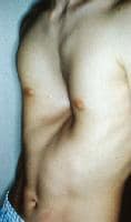
A 16-year-old boy with severe pectus excavatum. Note the appearance of the caved-in sternum and lower ribs.
Examples of pectus excavatum in young girls are shown in the images below.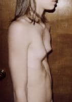
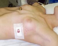
A 10-year-old girl with severe pectus excavatum. In girls, the deformity is of particular concern because of the medial displacement of the breast, resulting in significant asymmetry of the breasts and nipples (cross-eyed appearance of the nipples).
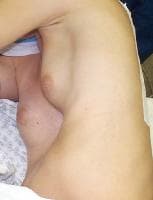
A 10-year-old girl with severe pectus excavatum. Note the significant asymmetry of the breasts and nipples (cross-eyed appearance of the nipples).
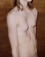
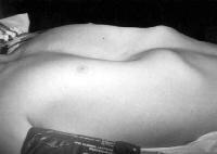
A 12-year-old girl with severe pectus excavatum. Note the significant asymmetry of the breasts. Preoperative photograph.
Pathophysiology
Frequency
United States
International
Mortality/Morbidity
Race
Sex
Age
Clinical
History
Physical
Causes
Other Problems to Be Considered
Workup
Laboratory Studies
Imaging Studies
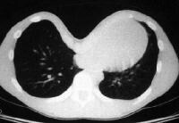
Preoperative CT scan of the chest of 12-year-old girl with severe pectus excavatum (see Media file 5). Note the severe pectus excavatum with compression of the lung fields and complete displacement of the heart and mediastinal structures to the left hemi-thorax.
Other Tests
Procedures
Histologic Findings
Treatment
Medical Care
Surgical Care
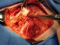
Operative photograph of the open Ravitch technique for repair of pectus excavatum. The anterior chest is exposed through an anterior thoracic incision and, after raising muscle and skin flaps, each involved cartilage is excised with preservation of the perichondrium. The picture shows one of the cartilages being removed.
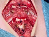
Operative photograph of the completed Ravitch procedure for correction of pectus excavatum. Note the sternum fractured at 2 different points with a cartilage graft in place to maintain its new position. The involved ribs underwent perichondrial excision. The deformity is completely corrected.
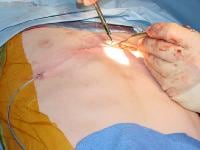
Closure of the anterior chest wall incision used for the open type of repair of pectus excavatum (Ravitch operation). Note the drain (small tubing) coming out on the side of the chest. Drains are typically removed after 2-3 days, and they prevent the accumulation of fluid under the skin and muscle flaps created at the time of surgery.
{{mediacaption:1005080_16}}
The image below is an illustration of another open operative technique used rarely today, the so-called "sternal turn-over operation."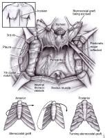
Operative diagram illustrating one of the open techniques for correction of pectus excavatum. The drawing is of the so-called "turn-over operation" for repair of pectus. It shows the extensive dissection and the radical nature of this open technique for surgical correction of this congenital chest wall deformity.
A 12-year-old girl with severe pectus excavatum. Note the significant asymmetry of the breasts. Preoperative photograph.
A 12-year-old girl with severe pectus excavatum immediately after minimally invasive repair. Note the immediate correction of the deformity.
Preoperative photograph of a 12-year-old boy prior to minimally invasive repair of pectus excavatum.
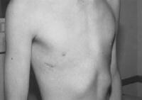
A 12-year-old boy 2 weeks after minimally invasive repair of his pectus excavatum. Note the small lateral chest wall incision and the excellent appearance of the anterior chest with 100% correction of the pectus deformity.
Indications for surgical repair
Operative correction should be considered in patients who present with pectus excavatum and cardiopulmonary impairment. The most common goal in operative repair of pectus excavatum is to correct the chest deformity. This is particularly important in teenagers, in whom the appearance of the chest can result in significant problems related to body image and self-esteem. Thus, the desire to improve the appearance of the chest is considered an appropriate medical indication for surgery. The images below illustrate the dramatic appearance of pectus excavatum in young male and female patients.A 16-year-old boy with severe pectus excavatum. Note the appearance of the caved-in sternum and lower ribs.
A 10-year-old girl with severe pectus excavatum. In girls, the deformity is of particular concern because of the medial displacement of the breast, resulting in significant asymmetry of the breasts and nipples (cross-eyed appearance of the nipples).
A 10-year-old girl with severe pectus excavatum. Note the significant asymmetry of the breasts and nipples (cross-eyed appearance of the nipples).
Other indications include exercise and physical activity limitations, evidence of cardiac or pulmonary dysfunction, chest pain, psychological distress, and potential future need for sternotomy (open-heart surgery). Adult patients with pectus excavatum who undergo open-heart surgery typically have significant displacement and rotation of the heart to the left chest. This can make the operative approach to the heart at the time of open-heart surgery difficult and challenging. With this in mind, elective repair of the pectus deformity prior to open-heart surgery may be indicated in selected cases.
Minimally invasive surgery for repair of pectus excavatum
First, children have a very soft and malleable chest. Second, the phenomenon of chest remodeling is well known in adult patients with emphysema who develop a barrel-shaped chest. If the chest wall in older adults can be reconfigured, the same should be possible in children and teenagers because of the increased malleability of their anterior chest wall. Third, the use of braces and internal fixating devices has allowed orthopedic surgeons and orthodontists to correct skeletal anomalies such as scoliosis, club foot, and maxillomandibular malocclusion. The anterior chest wall, which is quite malleable, is ideal for this type of correction.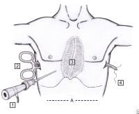
Illustration showing the minimally invasive technique for correction of pectus excavatum (3) with thoracoscopy (1). Note the long clamp passed from one side to the other (2) grabbing the umbilical tape (4), which serves as a guide for passage of the pectus bar behind the sternum.
Illustration showing the minimally invasive technique for correction of pectus excavatum (3) with thoracoscopy (1). Note the long clamp passed from one side to the other (2) grabbing the umbilical tape (4), which serves as a guide for passage of the pectus bar behind the sternum.
Illustration showing the minimally invasive technique for correction of pectus excavatum (3) with thoracoscopy (1). Note the long clamp passed from one side to the other (2) grabbing the umbilical tape (4), which serves as a guide for passage of the pectus bar behind the sternum.
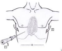
Operative diagram illustrating the pectus bar after it has been passed behind the sternum (5), under thoracoscopic visualization (1), before turning it over. Note that the concavity of the bar is facing up.
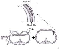
Illustration of the pectus bar passed behind the sternum before and after it is turned over. The insert shows the proper technique for fixation of the pectus bar against the lateral chest wall musculature.
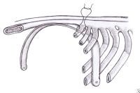
Illustration of the placement of the third point of fixation for stabilization of the pectus bar. Note that the nonabsorbable suture is placed around the bar and around a rib, lateral to the sternum on the anterior chest wall.
Critics of the MIRPE claim that the Nuss procedure is too invasive, risky, and not pain free. Proponents argue that this new approach, compared with the open surgery (modified Ravitch operation), eliminates the need for a large anterior chest wall incision with creation of pectoralis muscle flaps, resection of several ribs and cartilages, and sternal osteotomies. The MIRPE allows for a much shorter operating time, minimal blood loss, and minimal anterior chest wall scar. Moreover, the stability and strength of the chest wall is not compromised as it is with the open repair. For a more detailed review of the pros and cons of both approaches, please refer to the article "To Nuss or Not to Nuss? Two Opposing Views" in the Spring 2009 edition of Seminars of Thoracic and Cardiovascular Surgery.12
The current recommendations support the use of MIRPE in patients aged 5-20 years. The ideal age for undergoing this operation is 8-12 years because the chest wall is still very malleable, stabilization of the bar is easily achieved, thoracic epidural can be safely placed, and the child is mature enough to understand the operation and postoperative instructions, particularly incentive spirometry, which is essential for minimizing pulmonary problems after surgery.Consultations
Diet
Activity
Medication
Further Inpatient Care
Further Outpatient Care
Transfer
Deterrence/Prevention
Complications
Prognosis
Patient Education
Miscellaneous
Special Concerns
Multimedia
Media file 1: A 16-year-old boy with severe pectus excavatum. Note the appearance of the caved-in sternum and lower ribs. Media file 2: A 10-year-old girl with severe pectus excavatum. In girls, the deformity is of particular concern because of the medial displacement of the breast, resulting in significant asymmetry of the breasts and nipples (cross-eyed appearance of the nipples). Media file 3: A 10-year-old girl with severe pectus excavatum. Note the significant asymmetry of the breasts and nipples (cross-eyed appearance of the nipples). Media file 4: A 12-year-old girl with severe pectus excavatum. Note the significant asymmetry of the breasts. Preoperative photograph. Media file 5: A 12-year-old girl with severe pectus excavatum immediately after minimally invasive repair. Note the immediate correction of the deformity. Media file 6: Preoperative photograph of a 12-year-old boy prior to minimally invasive repair of pectus excavatum. Media file 7: A 12-year-old boy 2 weeks after minimally invasive repair of his pectus excavatum. Note the small lateral chest wall incision and the excellent appearance of the anterior chest with 100% correction of the pectus deformity. Media file 8: Preoperative CT scan of the chest of 12-year-old girl with severe pectus excavatum (see Media file 5). Note the severe pectus excavatum with compression of the lung fields and complete displacement of the heart and mediastinal structures to the left hemi-thorax. Media file 9: Illustration showing the minimally invasive technique for correction of pectus excavatum (3) with thoracoscopy (1). Note the long clamp passed from one side to the other (2) grabbing the umbilical tape (4), which serves as a guide for passage of the pectus bar behind the sternum. Media file 10: Operative diagram illustrating the pectus bar after it has been passed behind the sternum (5), under thoracoscopic visualization (1), before turning it over. Note that the concavity of the bar is facing up. Media file 11: Illustration of the pectus bar passed behind the sternum before and after it is turned over. The insert shows the proper technique for fixation of the pectus bar against the lateral chest wall musculature. Media file 12: Illustration of the placement of the third point of fixation for stabilization of the pectus bar. Note that the nonabsorbable suture is placed around the bar and around a rib, lateral to the sternum on the anterior chest wall. Media file 13: Operative diagram illustrating one of the open techniques for correction of pectus excavatum. The drawing is of the so-called "turn-over operation" for repair of pectus. It shows the extensive dissection and the radical nature of this open technique for surgical correction of this congenital chest wall deformity. Media file 14: Operative photograph of the open Ravitch technique for repair of pectus excavatum. The anterior chest is exposed through an anterior thoracic incision and, after raising muscle and skin flaps, each involved cartilage is excised with preservation of the perichondrium. The picture shows one of the cartilages being removed. Media file 15: Operative photograph of the completed Ravitch procedure for correction of pectus excavatum. Note the sternum fractured at 2 different points with a cartilage graft in place to maintain its new position. The involved ribs underwent perichondrial excision. The deformity is completely corrected. Media file 16: Closure of the anterior chest wall incision used for the open type of repair of pectus excavatum (Ravitch operation). Note the drain (small tubing) coming out on the side of the chest. Drains are typically removed after 2-3 days, and they prevent the accumulation of fluid under the skin and muscle flaps created at the time of surgery. 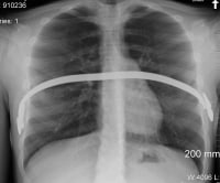
Media file 17: Chest radiograph of a 16-year-old patient in which the bar was displaced superiorly and the 2 stabilizers were separated from the Lorenz pectus bar as a result of intense physical activity during soccer practice. 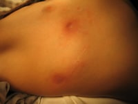
Media file 18: Skin rash secondary to a rare case of metal allergy caused by the pectus bar. 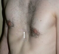
Media file 19: Recurrent pectus excavatum in a 24-year-old adult patient who underwent open repair using the Ravitch technique at age 10 years. 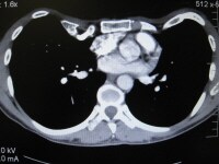
Media file 20: Chest CT scan of the recurrent pectus excavatum in the patient in Media file 20. 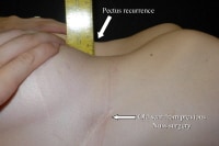
Media file 21: Recurrent pectus excavatum in young adult female patient who underwent minimally invasive repair at age 8 years. 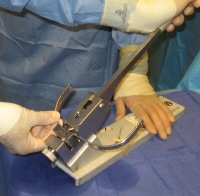
Media file 22: Technique for pectus bar bending. The Lorenz bar is bent by the operating surgeon at the time of pectus bar placement using an instrument known as the "bar bender." A smooth curvature is given to the bar so that it fits under the sternum and corrects the pectus deformity. 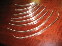
Media file 23: Pectus bars of various sizes. 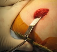
Media file 24: Technique for removal of the pectus bar. The bar and lateral stabilizer are easily exposed through the old lateral incision in the chest. Once exposed, it is pulled out using a bone-hook instrument. 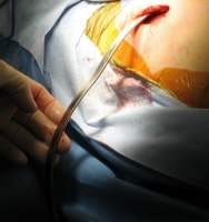
Media file 25: Technique for removal of the pectus bar. The bar is pulled out using a bone-hook instrument.
Saturday, March 27, 2010
(Enlarge Image)
(Enlarge Image)
(Enlarge Image)
(Enlarge Image)
(Enlarge Image)
(Enlarge Image)
(Enlarge Image)
(Enlarge Image)
(Enlarge Image)
(Enlarge Image)
(Enlarge Image)
(Enlarge Image)
(Enlarge Image)
(Enlarge Image)
(Enlarge Image)
(Enlarge Image)
(Enlarge Image)
(Enlarge Image)
(Enlarge Image)
(Enlarge Image)
(Enlarge Image)
(Enlarge Image)
(Enlarge Image)
(Enlarge Image)
Labels: Pectus Excavatum
Subscribe to:
Post Comments (Atom)
0 comments:
Post a Comment